Article co-authored by the Biomedical Society
Pupils in the Biomedical Society were truly inspired by our tour around the Peninsula Medical School laboratory in Derriford. The medical school runs in partnership with Plymouth and Exeter University and the NHS in Devon and Cornwall. Led by Dr Juri Na and Dr Leandro Jose De Assis, two specialist researchers working to reduce the growth of cancerous tumours, the tour focused on the main procedures used in their research. It’s interesting to note that whilst both doctors were working to improve the treatment of brain tumours, they were applying differing methods to tackle this problem. One focused on preventing transcription of DNA to stop the production of proteins that might make them more sensitive to radiotherapy, and the other, depriving the cancer cells of nutrition and slowing their metabolism and growth.
The tour started with a demonstration of two pupils working on cultures of immortalised schwannoma and meningioma cells. We learned how the Hayflick limit of cells in a sample can be exceeded by genetic modification. A cell will stop dividing when the telomeres that protect the chromosomes have fully eroded. Activation of an enzyme called telomerase prevents this gradual erosion of telomeres so cells can continue dividing, a key feature of many cancer cells. This means that cancer cell lines can continue to be studied for a longer period of time. In this room, the application of aseptic techniques was particularly evident and discussion ranged from cell regulation, Yellowstone Park geysers and Star Wars! (All scientifically relevant!) It was encouraging that despite complicated vocabulary and concepts, we were able to apply our A Level knowledge and engage genuinely with the staff.
Next, we were introduced to flow cytometry, a process which uses lasers and fluorescence (green fluorescent protein genetically modified into the cell DNA by inactivated HIV or using tagged antibodies) to analyse an entire liquid culture of cells. It was astounding to find that not only does a flow cytometer process 10,000 cells a second, the machine has the capacity to isolate a single cell using electromagnetic fields. This individual cell can then be cloned and tested on. We are planning to do a genetic modification practical involving GFP later this term so it was beneficial to see this be used in a practical way.
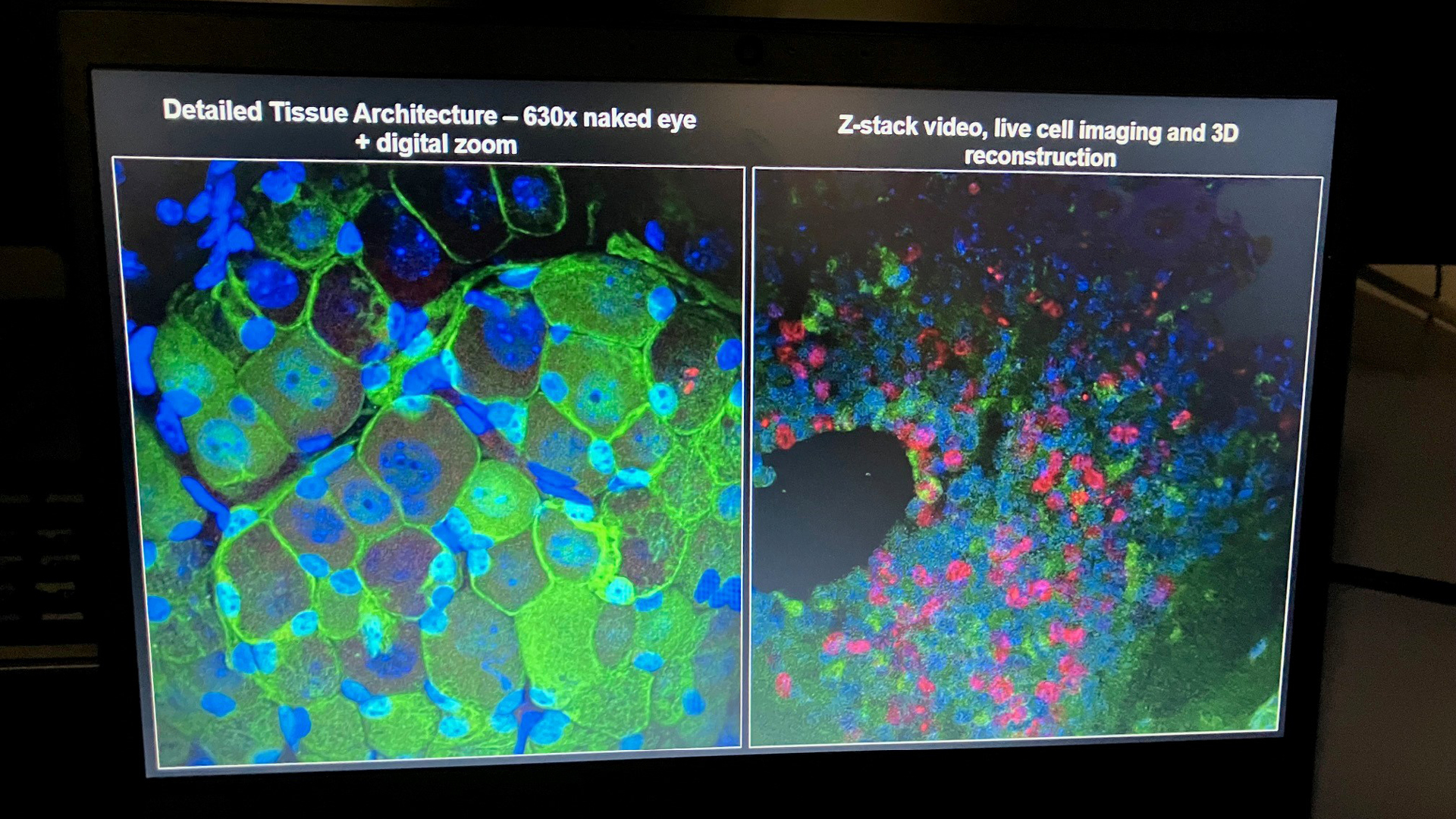
Seeing the images produced by a highly technical microscope was a great way to conclude the tour. Whether you enjoy science or not, the colourful pictures of tissue and cell imaging, will surely impress. The images are coloured based on properties of the cells using different light frequencies and tagged antibodies. Additionally, the microscope moves in and out of focus so that multiple layers of a sample can be compared to determine the prevalence of cancer cells.
The tour was an invaluable experience for those looking to work in the biomedical field and it was encouraging to contextualise what we’re learning at School. We’ve also gained a greater appreciation for the immense work that is put into the development for better medicine. Furthermore, the staff and pupils involved in the tour deserve a massive thank you for their enthusiasm and time, it was evident how much effort you had put into giving our class a great scientific experience! We would like to thank Dr Claire Adams and Dr Denis Wilkins especially for planning and organising such a wonderful tour.
Categories: Academic